What is immunophenotyping by flowcytometry?

immunophenotyping test
  • Flow cytometry is a laboratory method used to detect, identify, and count specific cells. This method can also identify particular components within cells.
  • This information is based on physical characteristics and/or markers called antigens on the cell surface or within cells that are unique to that cell type.
  • This method may be used to evaluate cells from blood, bone marrow, body fluids such as cerebrospinal fluid (CSF).
immunophenotyping test

Why Immunophenotyping by Flowcytometry:

Flow cytometry has been available for several decades and been adapted for use in many areas of clinical testing.

  • Sub-classification of leukemias and lymphoma
  • Myelodysplastic Syndrome (MDS) diagnosis
  • B-MRD (Minimal Residual Disease)- Identification of residual malignant B-Cells (blasts) in B-Cell Acute Lymphoblastic Leukemia after and during the course of treatment
  • T-MRD (Minimal Residual Disease)- Identification of residual malignant T-Cells (blasts) in T-Cell Acute Lymphoblastic Leukemia after and during the course of treatment
  • CD34 Stem Cell Enumeration (ISHAGE protocol)
  • High Sensitivity PNH Assay for diagnosis of paroxysmal nocturnal hemoglobinuria
  • Multiple Myeloma diagnosis and differentiation of neoplastic myeloma cells from reactive plasma cells
  • MM-MRD- Identification of residual malignant cells in multiple myeloma

Why MedGenome?

  • Unique form reporting as per CAP guidelines with cytochemistry examination images (MPO and PAS whenever is required) of blood/ bone marrow films
  • Scatter plots, analysed images and complete details of immunophenotyping so that the clinicians understands the case in detail
  • Use of new rare markers as LAIP (Leucocytes Aberrant Immunophenotype), theranostic and prognosis related markers in flow cytometry for future help especially during the course of treatment of patients

MedGenome Offer

Test Code

Test Name

Specimen Type

TAT

MGM997

CD34+ Stem cell Enumeration (CD45, CD34, 7AAD)

Bone marrow/ Peripheral blood in EDTA /Harvest Sample

4 hours

MGM1343

Leukemia/Lymphoma Panel- Flowcytometry

Peripheral blood along with bone marrow aspirate in EDTA

2 working days

MGM412

Acute Leukemia Classifier Panel – Flowcytometry

Peripheral blood along with bone marrow aspirate in EDTA, Peripheral blood can be accepted if blasts/atypical cells are high.

2 working days

MGM413

Acute Leukemia Screen Panel – Flowcytometry

Peripheral blood along with bone marrow aspirate in EDTA. Peripheral blood can be accepted if blasts/atypical cells are high.

2 working days

MGM414

Body fluid Leukemia/Lymphoma screen – Flowcytometry

Body fluid (effusions like CSF, pleural, ascitic, pericardial)

2 working days

MGM415

Chronic Lymphoid Leukemia Panel – Flowcytometry

Peripheral blood along with bone marrow aspirate in EDTA. Peripheral blood can be accepted if blasts/atypical cells are high.

2 working days

MGM417

Myelodysplasia Panel – Flowcytometry

Peripheral blood along with bone marrow aspirate in EDTA

2 working days

MGM416

Lymphoproliferative Disorder Classifier Panel – Flowcytometry

Peripheral blood along with bone marrow aspirate in EDTA. Peripheral blood can be accepted if blasts/atypical cells are high.

2 working days

MGM1230

B MRD

Bone marrow in EDTA

2 working days

MGM1370

PNH by FLAER- High Sensitivity

Peripheral blood in EDTA

2 working days

MGM1374

T- MRD

Bone marrow in EDTA

2 working days

MGM418

Myeloma Panel – Flowcytometry (CD45, CD38, CD19, CD56, CD138, kappa, lambda)

Peripheral blood along with bone marrow aspirate in EDTA

2 working days

Multiple Myeloma MRD (MM-MRD)

Bone marrow in EDTA

2 working days


How is it performed?

flowcytometry

FAQs

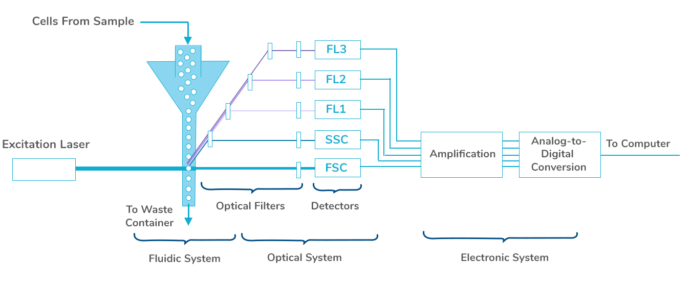
  • A sample of cells is suspended in a fluid.
  • Prior to testing and depending on the cells being analyzed, the sample may be treated with special dyes to further define cell sub-types. The dyes (fluorochromes) that are used are attached to monoclonal antibodies that bind to particular cells or key components of cells
  • The sample containing the cells passes through an instrument called a flow cytometer
  • In the instrument, the fluid in which the cells are suspended passes through very narrow channels so that the cells are organized in a single file as they pass the detector(s). This is accomplished at a high rate of speed (hundreds to thousands of cells per second.)
  • The flow cytometer contains one or more lasers and a series of photo detectors that are able to identify certain characteristics unique to various cell types. The single-cell suspension creates unique light-scattering events that occur when each cell passes through the laser light. These initial events are characteristic of the size and shape of the cell, as well as the intensity of the signal that is generated by the specific dyes, thus creating patterns that reflect cell type
  • The signals from the detectors are amplified and sent to a computer. They are converted to digital read-outs displayed on a computer screen or in a printout
  • The data are usually displayed as graphs

Free Genetic Counseling by MedGenome Genetic Experts

Please share your details with our genetic experts to answer your queries
Enquire Now Call Now

Contact Us

Business Hours: 8 AM - 8 PM

Toll-Free Hours: 9 AM - 7 PM

India:
MedGenome Labs Ltd.

3rd Floor, Narayana Nethralaya Building, Narayana
Health City, # 258/A, Bommasandra, Hosur Road
Bangalore – 560 099, India.
+91 80 67154932, +91 80 67154931

MedGenome Labs Ltd.

3rd Floor, Tower 1, E-City Software Park,
Electronic City – Phase 1, Bangalore – 560100, India.
+91 080-61171200

MedGenome Labs Ltd.

Second Floor, 11, Pusa Rd, Block 17 A, WEA,
Karol Bagh, New Delhi, Delhi 110005

US:
MedGenome Inc.

348 Hatch Dr, Foster City, CA 94404, United States

Download MedGenome Clinician App

© 2024 MedGenome . All Rights Reserved